María Jimena Costa
Ph.D. in Computer Science - Medical Image Processing
Segmentation of Anatomical Structures of the Lower Abdomen using 3D Deformable Surfaces.
Costa, J.
Ph.D. Thesis
Advisors: Prof. Ayache, N., Prof. Delingette, H.
École Nationale Supérieure des Mines de Paris - INRIA Project ASCLEPIOS
France, 2008
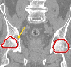
The main objective of this thesis is to provide radio--oncology specialists with automatic tools for delineating organs at risk of a patient undergoing a radiotherapy treatment of prostate tumors.
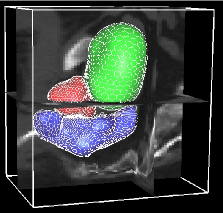
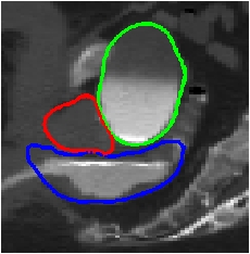
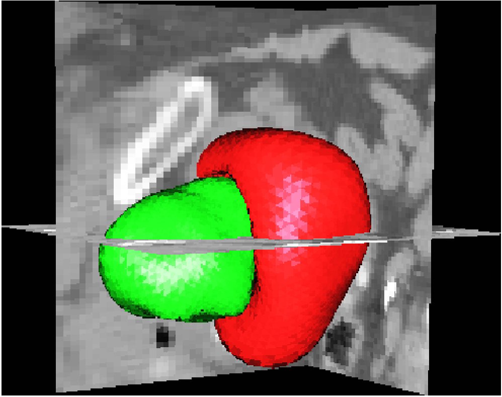
In order to achieve this goal, we work with CT Scan images. The images are first put in a common frame of reference by means of locally--affine registration based on the pelvic bone structures. A progressive approach consisting of three stages is then applied: the bladder is first delineated, the prostate is later included, and the rectum segmentation is finally integrated.
Given the highly heterogenous nature of the images in our database, our contribution for the segmentation process is centered on flexibility.
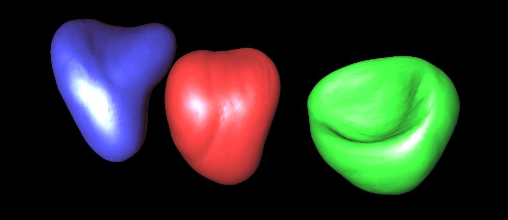
The bladder is a highly variable structure, both in terms of shape (fillings, compression by surrounding organs) and of intensity levels, the latter due to inhomogeneities caused by the presence or absence (to various levels) of a contrast agent. We propose a segmentation approach that is able to automatically adapt both to the shape and, most remarkably, to the intensity variability.
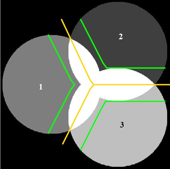
The prostate shows no distinct "edge" in the image itself; its interface with the bladder is often very difficult (if not impossible) to discern, even for the trained eye of medical experts. We have incorporated anatomical information and taken the intensity similarities into account in our approach to contour this structure. An original non--overlapping constraint optimizes the result in terms of image and shape prior information, in order to avoid ambiguities in the delineation of the common boundaries.
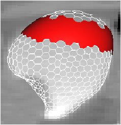
Finally, the rectum is incorporated in the segmentation. Different acquisition protocols for the CT scans result in images containing rectums with very different characteristics in terms of shape and intensity (due to filling level and natu re, air insufflation, contrast agent, etc.). A flexible method that makes no assumptions about the interior of the structure has been developped and thoroughly tested.
The developments that resulted from this thesis have been incorporated to the Isogray software by DOSIsoft, allowing further validation in clinical conditions.
Publications
Segmentation of Anatomical Structures of the Lower Abdomen using 3D Deformable Surfaces.
Costa, J.Ph.D. Thesis, March 2008
An Efficient Locally Affine Framework for the Smooth Registration of Anatomical Structures.
Commowick O., Arsigny V., Isambert A., Costa J., et al.Medical Image Analysis 2008
Automatic Segmentation of Bladder and Prostate Using Coupled 3D Deformable Models.
Costa J., et al.MICCAI 2007
Automatic Segmentation of the Bladder Using Deformable Models.
Costa, J., et al.ISBI 2007
An Efficient Locally Affine Framework for the Registration of Anatomical Structures.
Commowick O., Arsigny V., Costa J., Ayache N. and Malandain G.ISBI 2006
Towards an automatic delineation of lower abdomen structures for conformational radiotherapy based on CT images.
Costa J., et al.SFPM 2005.

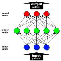
Distributed Ontological Encoding Through Symbol Recirculation.
Costa J., Duboue P.ASAI 2004.